Best Ultrasound Scan Test In Hyderabad-Trulife Diagnostics
Hyderabad, a thriving city in southern India, is well known for its state-of-the-art medical facilities and technology. Sonography scan tests are among the many diagnostic techniques that are accessible, and they are essential for identifying a variety of medical disorders. Sonography scans offer important insights into the internal structures of the body, and are used for everything from thorough abdominal examinations to frequent pregnancy checkups. This post explores the top Sonography scan tests, including cutting-edge sonography services, that Hyderabad has to offer.

What is an Ultrasound Scan?
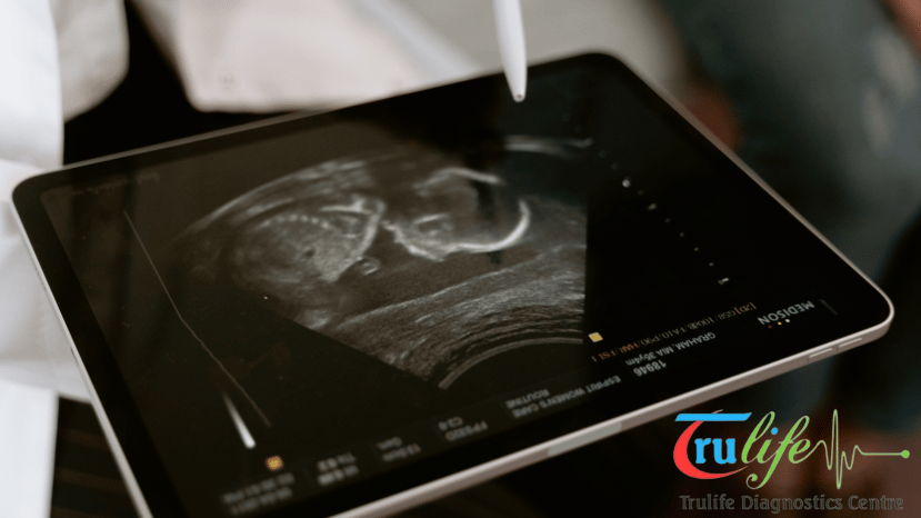
An Sonography scan is a type of imaging test that uses sound waves to create pictures of the inside of your body.
It is a safe and painless procedure that is often used to examine organs, muscles, and tissues. Trulife Diagnostic offers Sonography scans as part of their diagnostic services.
Sonography scans are used for a variety of purposes
- To monitor the development of a fetus during pregnancy
- To examine the organs in the abdomen, such as the liver, kidneys, and uterus
- To check for blood flow problems in the arms and legs
- To guide biopsies, which are procedures to remove a small sample of tissue for examination
- To diagnose tumors and other abnormalities
Why do I need an Ultrasound Scan?
Healthcare practitioners employ Ultrasonic Scans as a vital diagnostic tool to look at the body’s internal organs and tissues.
There are various reasons why you could require an Ultrasound Scan, whether you’re going through a standard screening or you’re exhibiting certain symptoms.
Here are some common scenarios where an Ultrasound Scan is recommended:-
Prenatal Care: Expectant mothers often undergo Ultrasound Scans during pregnancy to monitor the health and development of the fetus.
These scans can detect any abnormalities, assess fetal growth, determine the baby’s position, and even reveal the baby’s gender.
Abdominal Pain or Discomfort: If you’re experiencing unexplained abdominal pain, discomfort, or digestive issues, an abdominal Ultrasound can help identify the underlying cause.
It can visualize organs such as the liver, gallbladder, pancreas, kidneys, and spleen, providing valuable insights for diagnosis and treatment.
Pelvic Issues: For both men and women, pelvic Ultrasound Scans are commonly used to evaluate reproductive organs such as the uterus, ovaries, and prostate gland.
These scans can detect conditions like ovarian cysts, uterine fibroids, and prostate enlargement, aiding in the diagnosis of various gynecological and urological disorders.
Monitoring Medical Conditions: If you’ve been diagnosed with certain medical conditions such as heart disease, thyroid disorders, or kidney problems, regular ultrasound scans may be recommended to monitor the progression of the disease and assess treatment effectiveness.
Screening for Cancer: Ultrasound Scans can play a vital role in cancer screening and early detection.
Breast ultrasound, for example, is often used as a supplemental screening tool for women with dense breast tissue, helping detect breast cancer at an early stage when treatment is most effective.
Assessment of Soft Tissues and Musculoskeletal System: Ultrasound Scans can also be used to evaluate soft tissues, muscles, tendons, and joints for injuries, inflammation, or abnormalities.
Musculoskeletal Ultrasound is particularly useful for diagnosing conditions like tendonitis, bursitis, and ligament tears.

How Is The Ultrasound/Sonography Scan Test Is Performed?
- Preparation: Before the Ultrasound Scan, the expectant mother may be asked to drink water and have a full bladder, especially for early pregnancy scans or pelvic ultrasounds. This helps improve the visibility of the pelvic organs and the fetus. In some cases, specific preparations may be necessary based on the type of ultrasound being performed.
-
Positioning: The expectant mother will be asked to lie down on an examination table, usually in a comfortable position. Depending on the type of Ultrasound being performed, the sonographer may request different positions, such as lying on the back or side.
-
Gel Application: A clear water-based gel is applied to the skin over the area being examined. This gel helps facilitate the transmission of sound waves between the Ultrasound probe and the skin, ensuring clear images.
-
Probe Placement: The sonographer gently moves a handheld device called a transducer or probe over the skin in the area of interest. The probe emits high-frequency sound waves into the body, which bounce off internal structures and are then picked up by the probe. These sound waves are converted into real-time images on a monitor.
-
Image Acquisition: As the sonographer moves the probe, they carefully examine the images displayed on the monitor, capturing still images or video clips of specific structures or organs as needed. They may adjust the position of the probe or ask the patient to change positions to obtain optimal images from different angles.
-
Interpretation: The sonographer interprets the Ultrasound images in real-time, assessing the size, shape, and movement of organs, as well as the development and health of the fetus (in the case of prenatal ultrasounds). They may also take measurements of specific structures for diagnostic purposes.
-
Documentation: The findings from the Ultrasound examination are documented in a report, which may include images, measurements, and observations. This report is then reviewed by a radiologist or obstetrician-gynecologist, who may provide further interpretation and recommendations based on the results.

7 Reasons to get Ultrasounds Scan during pregnancy?
During pregnancy, Ultrasound Scans are essential for monitoring the health and development of both the mother and the fetus.
Trulife Diagnostic, a renowned healthcare facility in Hyderabad, offers comprehensive prenatal ultrasound services to ensure optimal pregnancy care.
Here are some key reasons why getting Ultrasounds during pregnancy is crucial:
- Confirmation of Pregnancy: An ultrasound scan is often the first step in confirming a pregnancy. By visualizing the gestational sac and fetal heartbeat, ultrasound helps verify the presence of a viable pregnancy, providing reassurance to expectant parents.
- Dating the Pregnancy: Ultrasound can accurately determine the gestational age of the fetus, helping healthcare providers establish an estimated due date. This information is vital for monitoring fetal growth and development throughout the pregnancy.
- Assessment of Fetal Growth: Regular ultrasound scans allow healthcare providers to monitor the growth of the fetus and ensure that it is developing at a healthy rate. Abnormalities in fetal growth can indicate potential complications such as intrauterine growth restriction (IUGR) or macrosomia, which may require further intervention.
- Detection of Fetal Anomalies: Ultrasound Scan can detect structural abnormalities or congenital anomalies in the fetus, such as neural tube defects, heart defects, or limb abnormalities. Early detection of these conditions enables parents to make informed decisions about their pregnancy and plan for any necessary medical interventions or treatments.
- Evaluation of Placental Health: Ultrasound can assess the position and health of the placenta, ensuring that it is properly attached to the uterine wall and functioning correctly. Abnormalities in placental location or function, such as placenta previa or placental insufficiency, can increase the risk of complications during pregnancy and childbirth.
- Monitoring Amniotic Fluid Levels: Ultrasound can measure the volume of amniotic fluid surrounding the fetus, ensuring that it remains at appropriate levels throughout pregnancy. Abnormalities in amniotic fluid volume, such as oligohydramnios or polyhydramnios, may indicate underlying fetal or placental issues that require further evaluation and management.
- Guidance for Prenatal Interventions: In certain cases, ultrasound-guided procedures may be necessary during pregnancy, such as amniocentesis or chorionic villus sampling (CVS) for genetic testing. Ultrasound provides real-time imaging to guide these interventions safely and accurately, minimizing the risk to both the mother and the fetus.
By offering advanced ultrasound technology and experienced sonographers, Trulife Diagnostic ensures that expectant mothers in Hyderabad receive comprehensive prenatal care. From early pregnancy confirmation to monitoring fetal growth and detecting potential complications, Ultrasound Scans play a crucial role in ensuring the health and well-being of both mother and baby throughout the pregnancy journey.
What are the different types of Ultrasound Scan for Pregnancy?
During pregnancy, various types of ultrasound scans are performed to monitor the health and development of the fetus. Trulife Diagnostic, a leading healthcare facility in Hyderabad, offers a comprehensive range of ultrasound services tailored to the specific needs of expectant mothers. Here are the different types of ultrasound scans commonly used during pregnancy:
- Dating Scan: This ultrasound scan is typically performed in the first trimester, usually between 8 to 14 weeks of pregnancy. It is used to accurately determine the gestational age of the fetus and establish an estimated due date. The dating scan also confirms the number of fetuses (single or multiple pregnancies) and assesses the viability of the pregnancy.
- Nuchal Translucency (NT) Scan: The NT scan is usually performed between 11 to 14 weeks of pregnancy. It measures the thickness of the fluid at the back of the baby’s neck to assess the risk of chromosomal abnormalities, particularly Down syndrome. Combined with maternal blood tests, the NT scan provides valuable information for early prenatal screening.
- Anomaly Scan (Level II Scan): Also known as the mid-pregnancy ultrasound or fetal anomaly scan, this comprehensive scan is typically performed between 18 to 22 weeks of pregnancy. It evaluates the fetal anatomy in detail, assessing the development of organs, limbs, spine, and facial features. The anomaly scan helps detect structural abnormalities or congenital anomalies in the fetus.
- Fetal Growth Scan: Fetal growth scans, also called growth ultrasounds or growth scans, are performed periodically throughout the pregnancy to monitor the growth and development of the fetus. These scans assess parameters such as fetal size, weight, and abdominal circumference, ensuring that the baby is growing at an appropriate rate for its gestational age.
- Doppler Ultrasound: Doppler ultrasound measures the blood flow in the umbilical cord, placenta, and fetal blood vessels. It helps assess placental function, detect abnormalities in blood circulation, and monitor fetal well-being, particularly in high-risk pregnancies or cases of suspected fetal growth restriction.
- Biophysical Profile (BPP): The biophysical profile combines ultrasound imaging with fetal heart rate monitoring to assess fetal well-being. It evaluates fetal movements, breathing movements, muscle tone, amniotic fluid volume, and fetal heart rate patterns. The BPP is often recommended in cases of high-risk pregnancies or concerns about fetal health.
- 3D/4D Ultrasound: While not essential for medical diagnosis, 3D and 4D ultrasound scans provide three-dimensional and real-time images of the fetus, allowing expectant parents to see detailed facial features and movements. Trulife Diagnostic may offer these optional scans for bonding purposes and a more immersive prenatal experience.
By offering a range of ultrasound scans tailored to different stages of pregnancy, Trulife Diagnostic ensures comprehensive prenatal care and early detection of any potential issues. These ultrasound scans play a crucial role in monitoring fetal health, guiding medical management, and providing peace of mind to expectant parents throughout their pregnancy journey.
Different types of Sonography Scan for Pregnancy are:

Fetal echocardiography (fetal echo): A test that uses sound waves (Sonography) to produce images of the baby’s heart.
This test examines the developing baby’s heart walls and valves, the blood vessels leading to and from the heart, and the heart’s pumping strength.
Three-dimensional (3-D) Sonography – 3-D Sonography for Pregnancy generates images that look more lifelike than standard Sonography images. 3D scans show still pictures of the baby in three dimensions.
These USG images can give doctors and radiologists a better picture of the baby’s development inside the womb.
Four-dimensional (4-D) Sonography– 4-D Sonography for Pregnancy is similar to a 3D Sonography, but the picture shows movement like a video.3D and 4D Sonography scans may easily find fetal abnormalities.
These scans can display more useful information from different angles, for example, cleft lip.
This information can help doctors to decide on a treatment plan for a baby’s cleft lip after birth.
Transvaginal ultrasound: The radiologist keeps a transducer inside the vagina. This test is usually done during the early stage of Pregnancy when the uterus is still small and close to the vagina.
Doppler Sonography: This test is used to check the blood flow in the baby’s heart, through the umbilical cord, and between the baby and the placenta.
Doppler imaging can also be used to check a mother’s blood pressure and examine if the baby is growing normally.


